Microglia are the tissue macrophages of the brain, cleaning up cellular debris and constantly surveilling their environment. They form branches called ramifications with which they reach out to specific parts of the neurons, such as the axon initial segment. Viktor al-Naqib, neurobiologist in the Axonal Signaling Group of Maarten Kole at the Netherlands Institute for Neuroscience in Amsterdam, set out to study these interactions, because as he says: "We don't know why and under what circumstances the microglia are attracted to the axon initial segment."
But microglia are very difficult to study, found Viktor. "Normally, we study neurons in a slice of brain tissue. But microglia, being partially responsible for garbage disposal in the brain, become too activated and scavenge the damaged brain tissue."
The solution is working with brain slice cultures, he explains. "The difference is the time scale. We now have days or even weeks to study the microglia. Right after slice preparation, they are still very activated, but eventually they chill out and go into their homeostatic state. Then we can observe what they do."
Photocages
In this ICI-project, the aim is to develop a toolkit to study these complex cells, both by manipulating the immunological state of the microglia and studying the influence of certain molecules such as ATP that are known to be involved in their function.
In the Molecular Immunology group of prof. Sander van Kasteren at Leiden University, PhD student Yixuan Wang designed and synthesized so-called photocaged compounds. These compounds are inactive as long as they are bound to a protective group. This group is photolabile and can be removed by shining specific light on it.
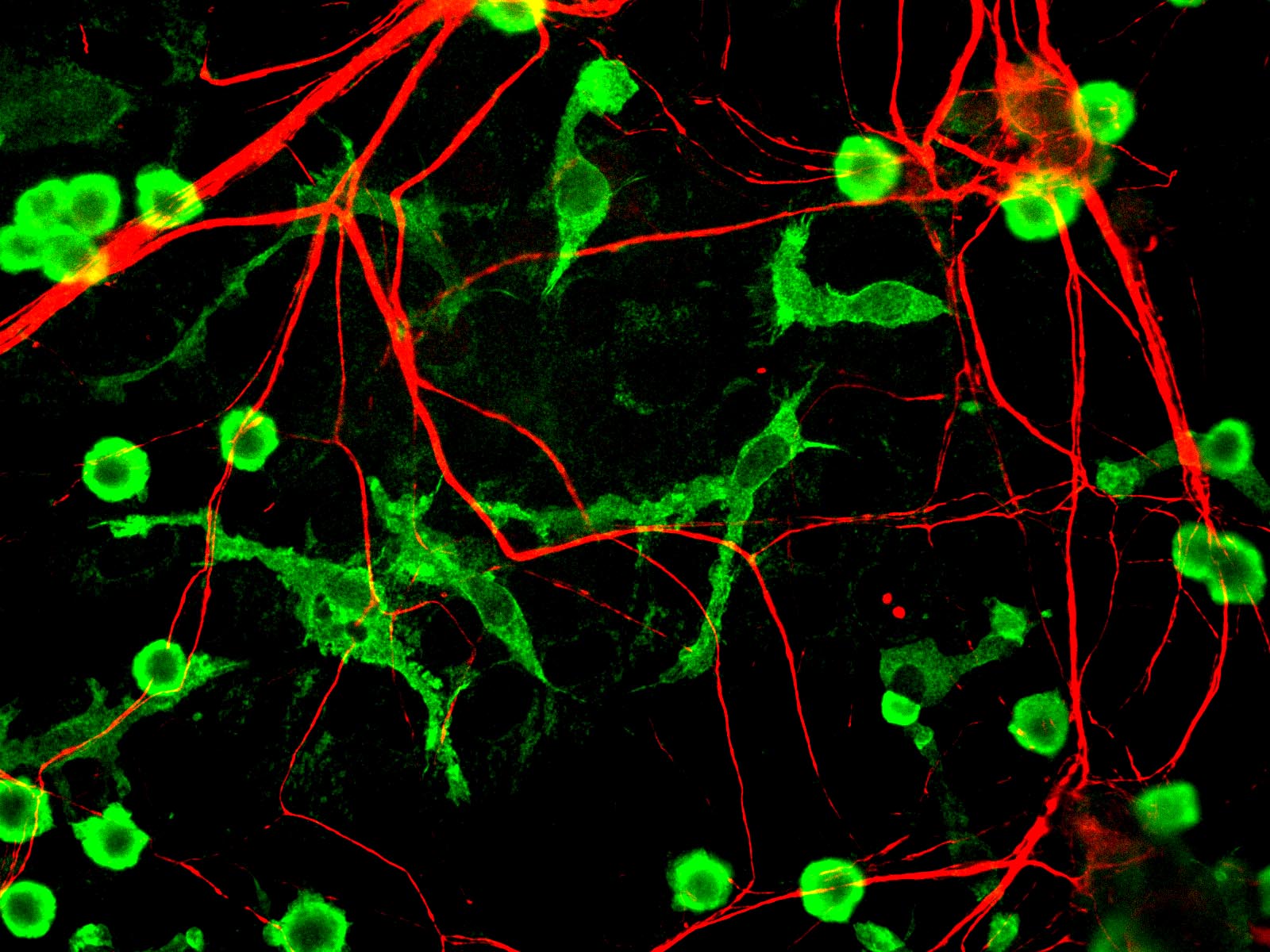
For Viktor, this means he can prepare a brain cell culture with microglia he wants to study, using fluorescent reporter lines so they are visible under a microscope. He then adds the photocaged ATP (or other photocaged compounds) and specifically activates it near one microglia cell. He can then study what happens. In the case of photocaged ATP he expects the microglia to become attracted to the activated, uncaged area, but he can also study what happens if he adds an inhibitor of ATP receptors, or when all the neurons around the microglia stop communicating, for example.
"Although the experiments with the photocaged ATP are not fully working yet, I am happy that we now have a nicely working protocol to study the activity of microglia and the axon in detail. When the compounds will work they will be amazing tools to study microglial biology in a highly targeted manner. Once we have a better fundamental understanding of how microglia are activated and how they talk to neurons, we can start to work towards an understanding of neurological diseases associated with them."
Close cooperation
Working in such close cooperation in a multidisciplinary team has been very exciting, says Viktor. "We have frequent meetings where we get ideas and suggestions. I get to observe how they do problem solving. We work at the interface of very different disciplines but we have a shared language which is science."
Project: The Development Of Photochemical Tools For In Vivo Microglial Activation To Control And Study Microglia-Neuron Crosstalk In Space And Time.
Viktor al-Naqib, Netherlands Institute for Neuroscience, Axonal signaling group of Maarten Kole.
This article was published in ICI Bulletin 16, december 2023.